
World-Leading Microscope Shows More Detail Than Ever
Posted on: May 8, 2006
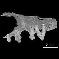
Photo Courtesy: EPSRC
A unique 3-dimensional microscope that works in a new way is giving unprecedented insight into microscopic internal structure and chemical composition. It is revealing how materials are affected, over time, by changes in temperature, humidity, weight load and other conditions.
The device could lead to advances in a range of areas, such as healthcare (in furthering, for instance, the understanding of conditions such as osteoporosis), the development of better construction materials, improved oil extraction methods and even the study of fossils.
Like a number of other microscopes, the new microscope harnesses X-rays to provide information about an object’s internal structure down to micron scale. (A micron is a millionth of a metre.) What makes it unique, however, is its innovative use of a technique called ‘time delay integration’, which enables it to generate much better images of larger objects than any other device. This means that microscopic structure can be studied with greater accuracy.
With EPSRC funding, a multi-disciplinary team drawn from six UK universities has been developing and utilising the microscope, which, although similar to the CT scanners used in healthcare, can view things in much greater detail.
X-ray microscopes can produce 3-d internal pictures of an object by taking a large number of 2-d images from different angles – this is known as X-ray microtomography. However, the new microscope’s combining of this technique with time delay integration is completely unique. Through averaging out imperfections in the image across all pixels, this approach enables the microscope to produce clearer and bigger pictures than previously possible (see ‘Notes for Editors’).
Some of the microscope’s many potential uses include:
- Studying how bone and tooth tissue behave in conditions such as osteoporosis, osteoarthritis and tooth decay. By improving understanding of these conditions, the microscope could aid prevention, earlier diagnosis and more effective treatment.
- Observing how crude oil is held in sandstone pores. This knowledge could assist the development of more efficient ways of exploiting both offshore and onshore oil resources.
- Investigating the mechanical behaviour of metals at a microscopic level. This could contribute to development of more reliable, more resilient and lower weight materials for use in construction, aviation and the storage & transportation of dangerous substances.
- Detailed study of fossils embedded in rocks without having to remove and risk damaging them.
The microscope looks set to be a valuable research tool that many different organisations in a wide range of sectors could benefit from using. The team is currently planning to seek funding to support the development of a radical new design that could be even more effective.